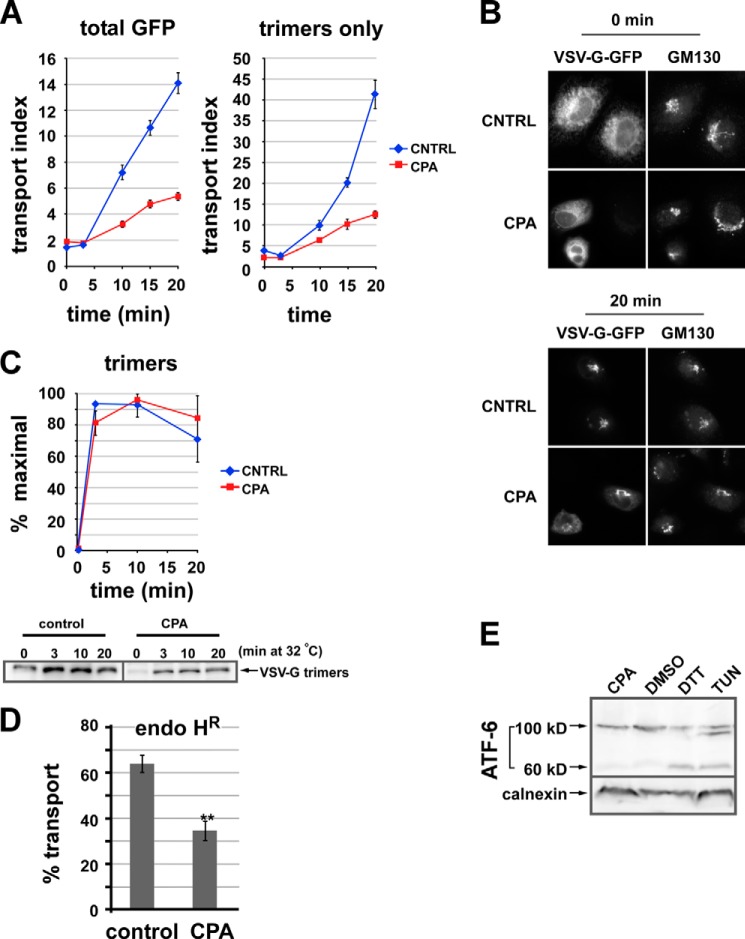
FIGURE 1.
Luminal calcium depletion inhibits ER-to-Golgi transport of VSV-G-GFP trimers. A, VSV-G-GFP-expressing NRK cells were CPA- or mock-treated. Cargo was then released from the ER by shifting cells from 40 to 32 °C. Cells were fixed after 0, 3, 10, 15, or 20 min; permeabilized; and labeled to detect VSV-G trimers using the I-14 antibody. The arrival of total VSV-G-GFP fluorescence (left plot) or VSV-G trimers (right panel) in the Golgi was quantified. Each value is a mean of ≥20 cells from a single experiment repeated at least three times with a similar outcome. Error bars, S.E. The transport index is much higher in the right panel because unfolded VSV-G-GFP in the ER is not detected. B, representative images from the experiment quantified in A. C, VSV-G trimerizes in 3 min. Cells expressing VSV-G-Myc or VSV-G-GFP were CPA- or mock-treated prior to temperature shifts and then chilled and lysed. Assembled VSV-G trimers were immunoprecipitated using the I-14 antibody and detected by immunoblot. The graph (top) was generated by averaging three experiments. Blots from one of the experiments are shown (bottom). D, CPA treatment inhibits ER-to-Golgi transport measured by acquisition of endoglycosidase H resistance (endo HR). NRK cells were transfected with VSV-G-Myc ts045 and grown for 24 h at 40 °C and then either left in regular medium (control) or else treated with CPA as in Fig. 1 at 40 °C (CPA). Cells were then shifted to 32 °C for 0 or 40 min to allow transport through the medial Golgi prior to standard procedures for determination of endo H resistance using endo H digestion followed by SDS-PAGE and anti-Myc immunoblotting. Plotted is the percentage transport after 40 min at 32 °C, quantitated from three replicates with the formula, percentage transport = endo H-resistant/(endo H-resistant + endo H-sensitive) × 100%. The 0-min background value of percentage transport for cells left at 40 °C was subtracted for each group of cells (∼20% for CPA-treated cells). **, p < 0.01 in a two-tailed t test. E, ATF-6 is not activated by our Ca2+ depletion conditions. NRK cells were transfected with FLAG-ATF-6, treated, lysed, and immunoblotted to detect FLAG. Lane 1, CPA as in A; lane 2, DMSO for 2 h; lane 3, 1 mm DTT for 2 h; lane 4, 2 μg/ml tunicamycin (Tun) for 2 h. Uncleaved ATF-6 is 100 kDa, whereas the cleaved ATF-6 is ∼60 kDa.

