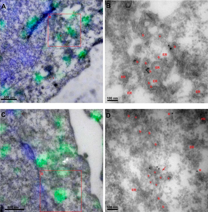
FIGURE 4.
Enlarged peripheral puncta induced by CPA contain abundant COPII buds and clusters of unfused vesicles. Correlative section light and electron microscopy of CPA structures in peripheral regions lacking Golgi. Anti-Sec31 primary antibody was followed by anti-rabbit Alexafluor-488 (green), 10-nm protein A-gold, and Hoechst (blue). Sections were imaged by fluorescence microscopy and then stained with uranyl acetate and embedded with methylcellulose prior to electron microscopy and fluorescence-transmission EM correlation. A and C, overlays of fluorescence with morphology visible by EM. Gold particles were enhanced with red. White arrowheads highlight swaths of fluorescence labeling over buds and vesicles not labeled by gold. B and D, magnified view of regions in red boxes in A and C, with buds (b), vesicles (v), VTCs, and ER labeled. Red arrow, vesicle cluster. Asterisks, gold particles not clearly associated with a membrane.

