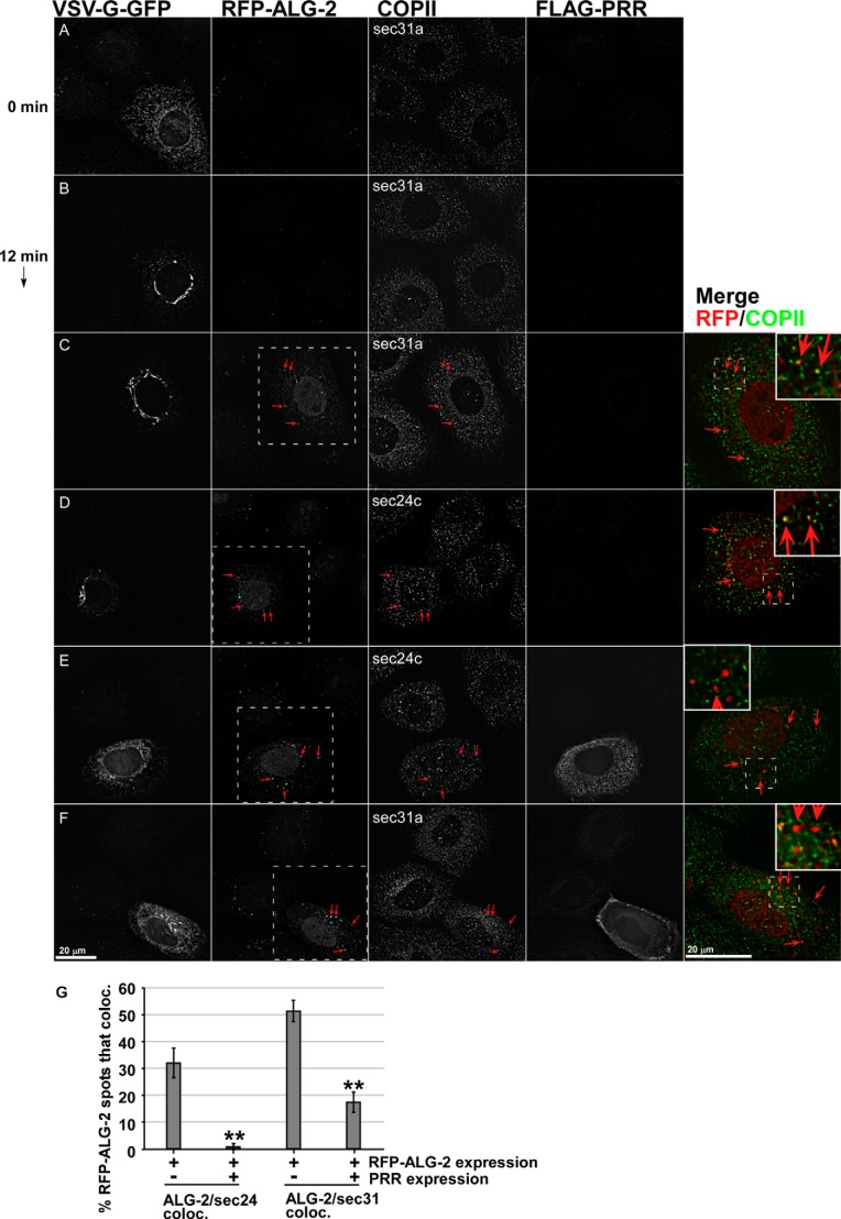
FIGURE 6.
Effects of ALG-2-RFP and Sec31A-PRR-FLAG on endogenous COPII components. NRK cells transfected with VSV-G-GFP along with no additional construct (A and B), with ALG-2-RFP (C and D), or with both ALG-2-RFP and FLAG-Sec31-PRR (E and F) were shifted to 32 °C for 0 (A) or 12 min (B–F) and then fixed and immunolabeled to detect FLAG-PRR and endogenous Sec31A (A–C and F) or endogenous Sec24C (D and E). Shown are single optical sections of deconvolved wide field image stacks. Arrows, prominent cytoplasmic puncta decorated by ALG-2-RFP that frequently co-localize with ERES and ERGIC markers. Merged images show superposition of the ALG-2-RFP (red) and COPII marker (green) in a magnified single cell from the field (marked with white boxes in the RFP-ALG-2 image). Insets within the merged planes show a smaller area with a higher magnification. G, quantification of the percentage of cytoplasmic ALG-2-RFP-positive structures that co-localize significantly with Sec24C and Sec31A. Error bars, S.E.

