FIGURE 1.

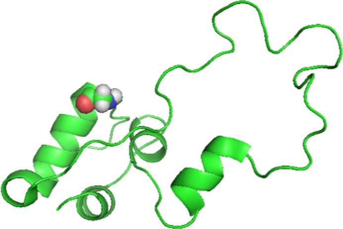
Structure of the proinsulin molecule as a schematic model with GlyB8 in the β-turn at amino acid residues B7-B10 (turn 1) shown with CPK (Corey-Pauling-Koltun) space-filling rendering. Image adapted from PDB code 2KQP.

Structure of the proinsulin molecule as a schematic model with GlyB8 in the β-turn at amino acid residues B7-B10 (turn 1) shown with CPK (Corey-Pauling-Koltun) space-filling rendering. Image adapted from PDB code 2KQP.