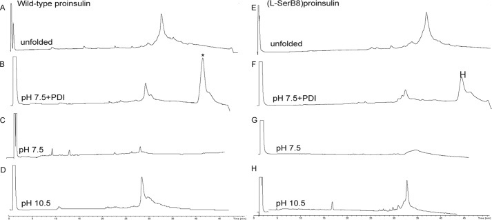
FIGURE 4.
Folding of wild-type proinsulin (left column) and [l-SerB8]proinsulin (right column) in three different conditions: at pH 10.5 (50 mm glycine/NaOH, 1 mm EDTA, 1 mm reduced GSH, 1 mm GSSG, at 4 °C and peptide concentration of 0.1 mg/ml); at pH 7.5 (10 mm Tris, 10 mm glycine, 1 mm EDTA, 1 mm GSH, 2 mm GSSG, 70 mm GdnHCl, at room temperature and peptide concentration of 0.1 mg/ml); and at pH 7.5 in the presence of PDI. The top two chromatograms represent the unfolded polypeptide of wild-type proinsulin (A) and [l-SerB8]proinsulin (E). The six bottom chromatograms represent the reaction mixtures after overnight folding under the conditions shown on each chromatogram. The asterisk (*) corresponds to the PDI protein. All the reactions were monitored by LC-MS and the masses were confirmed by online ESI.

