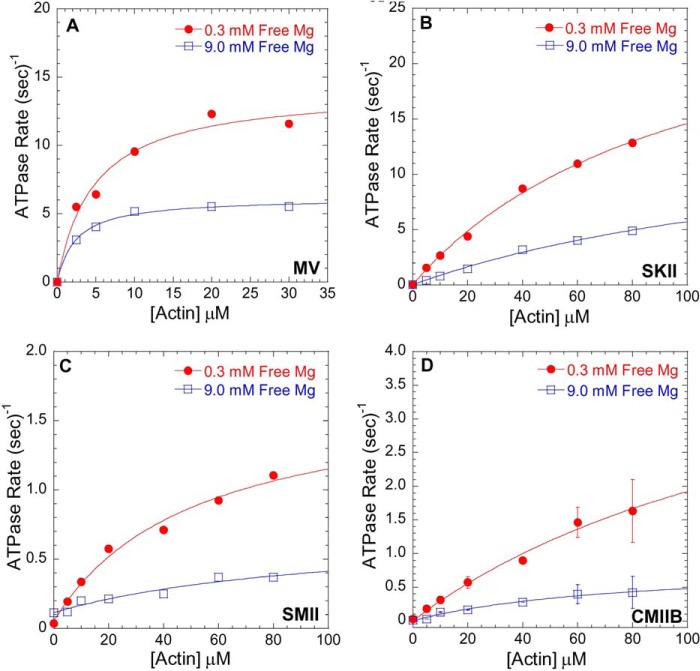
FIGURE 1.
Mg2+-dependent actin-activated ATPase activity. ATPase assays were performed in ionic strength controlled conditions with 0.3 mm free Mg2+ (filled red circles) or 9.0 mm free Mg2+ (open blue squares) for MV (A), SKII (B), SMII (C), and CMIIB (D) in the presence of different actin concentrations at 25 °C. See Table 1 for a summary of kcat and KATPase values.

