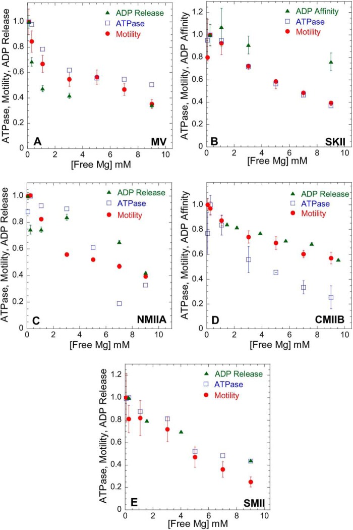
FIGURE 4.
Influence of Mg2+ on actomyosin ATPase, motility, and ADP release/affinity. Steady state ATPase (open blue squares), motility (filled red circles), and ADP release/affinity (filled green triangles) were performed in ionic strength-controlled conditions with 0.3–9.0 mm free Mg for MV (A) and members of the myosin II family as follows: SKII (B); NMIIA (C); CMIIB (D); and SMII (E). The ATPase assays were performed at 25 °C with fixed actin concentrations in MV (20 μm actin) and MII (60 μm actin). Actomyosin motility was performed at 27 °C (see Table 4 for velocity values). The data are expressed as relative values for comparison (normalized to the value obtained at 0.3 mm free Mg). The ADP release rate constant was examined with mant-dADP or pyrene actin (see Fig. 2), and ADP affinity was measured by competition with ATP-induced dissociation (see Fig. 3).

