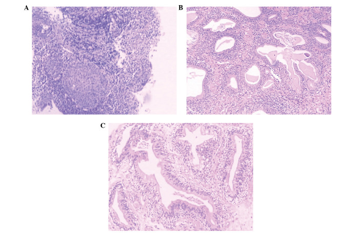
Figure 1.
(A and B) Typical cystitis glandularis. Urothelium show reactive changes and underlying proliferation of von Brunn’s nests. (C) Intestinal cystitis glandularis. Sections show the presence of goblet cells and a morphological similiarity to colonic mucosa (stain, trypan blue; magnification, ×100).

