
Figure 4.
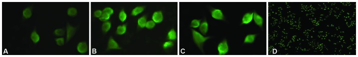
Experimental result of the immunofluorescence staining. The CDK6 protein is shown as bright green fluorescence, as observed by fluorescence microscopy using a wavelength of 475nm. (A) MGC-803 cells of the miR-449a upregulation group (magnification, ×400). (B) MGC-803 cells of the miR-449a downregulation group (magnification, ×400). (C and D) Cells of the control group. miR, microRNA; CDK6, cyclin-dependent kinase 6 (magnifications, ×400 for A and ×10 for D).