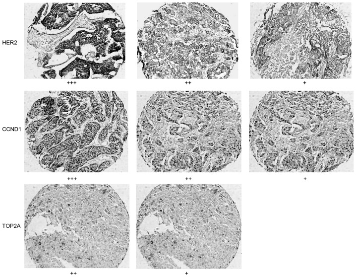
Figure 1.
Immunohistochemistry of HER2, CCND1 and TOP2A in patients with lymph node negative breast cancer with a good and poor prognosis. Imaging was performed at ×200 magnification. HER2+, weak and incomplete membrane staining in >10% of cells; HER2++, moderate and complete membrane staining in >10% of tumor cells; HER2+++, strong and complete membrane staining in >10% of tumor cells. CCND1+ and TOP2A+, positive cells were 10–20%; CCND1+ and TOP2A++, positive cells were 20–50% ; CCND1+ and TOP2A+++, positive cells were >50%.

