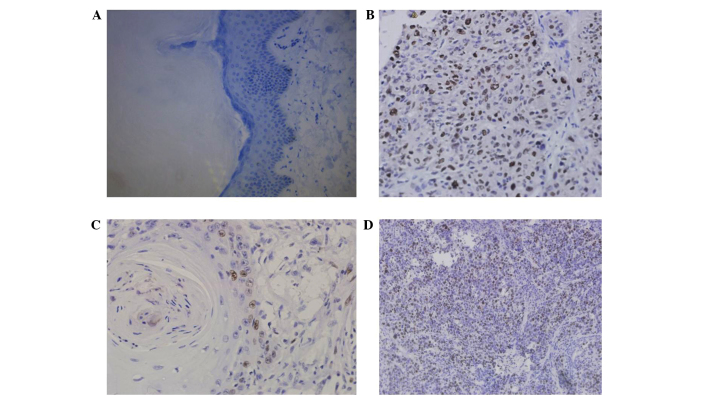
Figure 1.
Immunohistochemical detection of Topo II-α protein expression in laryngeal cancer tissue specimens compared with distant healthy tissues. (A) Healthy tissue without Topo II-α expression. (B) Laryngeal cancer tissue with Topo II-α expression (tumor cell nuclei appeared brown and yellow in color). (C) Well-differentiated laryngeal cancer. (D) Poorly differentiated laryngeal cancer. (Magnification: A, ×400; B, ×400; C, ×400; and D, ×200).

