Figure 6.
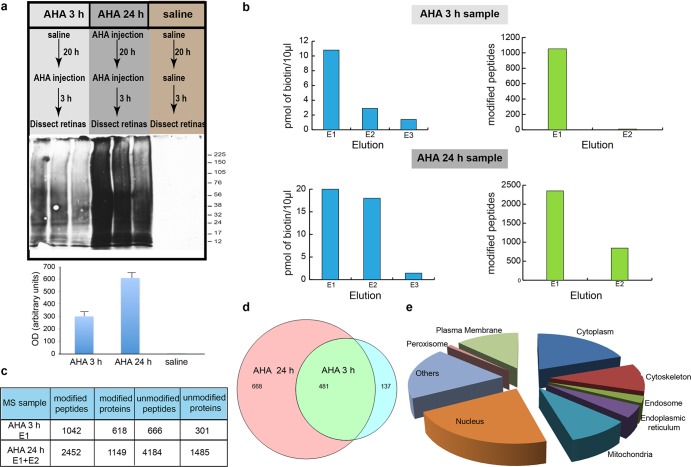
DiDBiT detection of newly synthesized proteins in adult rat retina in vivo. (a) Upper panel: Protocols for intraocular AHA injections to evaluate the temporal resolution of in vivo AHA labeling to detect de novo protein synthesis in the retina. Adult rats received intravitreal AHA injections and retinas were collected after 3 h (labeled “AHA 3 h sample”). Another group of animals received two intravitreal AHA injections 21 h apart and were sacrificed 3 h after the second injection (labeled “AHA 24 h sample”). AHA-labeled proteins were biotinylated by click chemistry and analyzed using DiDBiT. (a) Center and lower panels: Western blots and quantification of AHA-biotin labeled retinal proteins after click reaction with biotin-alkyne. More AHA-biotin labeled proteins are detected after 24 h of AHA labeling compared to 3 h. No biotin label is detected in samples from control animals after intravitreal injection of saline. (b) Biotin measurements (left panels) and MS detection of biotin-modified peptides (right panels) from sequential NeurAvidin elutions (E1–E3) of peptides from in vivo AHA labeling of newly synthesized proteins in the retina, analyzed by MudPIT. In the AHA 3 h sample, only E1 had sufficient biotin content to warrant MS analyses, whereas the two first elutions (E1 and E2) from the AHA 24 h sample group had sufficient biotin for MS analysis. (c) Numbers of modified and unmodified peptides and proteins from the 3 and 24 h retinal AHA samples. More unmodified proteins were detected in the 24 h AHA retina sample than the 3 h AHA retina sample because the sample was the combination of E1 and E2, both of which include unmodified and modified proteins. (d) Venn diagram showing numbers and overlap of newly synthesized proteins based on direct detection of AHA-biotin modified peptides after 3 and 24 h of AHA labeling. The majority (78%) of newly synthesized retinal proteins detected after the 3 h AHA labeling period were also detected after 24 h of AHA labeling. (e) Distribution of AHA-biotin labeled peptides and corresponding proteins in cellular compartments from the AHA 3 h sample. The 24 h labeling group resulted in the same cellular distribution of newly synthesized proteins (see Supporting Information Table 1).

