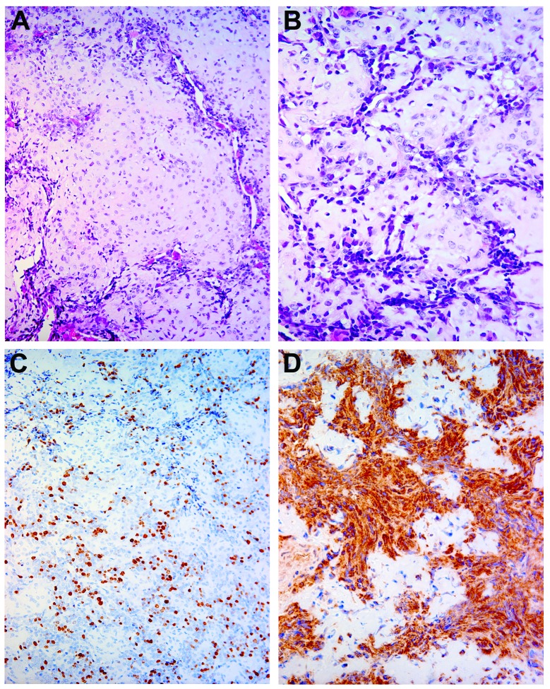
Figure 1.
(A and B) Typical morphological features of mesenchymal chondrosarcomas showing a biphasic pattern of cartilage islands distributed among spindle cells, mainly located in the periphery. Chondrocytes showed moderate nuclear atypia, while spindle cells exhibited nuclear hyperchromatism and pleomorphism [staining, haematoxylin and eosin; magnification, ×200 (A) and ×400 (B)]. (C) The proliferation index (Ki-67) was high in the spindle cell component, but low in the cartilage islands (magnification, ×200). (D) After staining for CD99 (MIC 2), strong immunoreactivity was observed only in the peripheral cellular part of the tumour (magnification, ×400).

