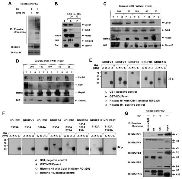
Figure 3. Mitochondrial CyclinB1/Cdk1 localizes in the Matrix and Phosphorylate CI Subunits at G2/M Transition.
(A) Immunoblotting analysis of mitochondrial (Mi) phospho-serine/threonine proteins isolated from G0/G1 (0 h) and G2/M(32 h) cells after release from G0/G1 synchronization.
(B) Sub-mitochondrial localization of CyclinB1 and Cdk1 detected by alkaline extraction (Antonyuk et al.). Matrix proteins were separated from integral membrane proteins by extracting mitochondria with sodium carbonate (pH 11), then the total input (T), soluble matrix proteins (S), and membrane vesicle pellets (P) were immunoblotted for CyclinB1, Cdk1, Tom40 (an outer membrane protein), TIMM13 (an inter-space protein), and HSP60 (a matrix protein).
(C, D) Sub-mitochondrial localization of CyclinB1 and Cdk1 detected via mitoplasting and protease protection assay (Antonyuk et al.). Mitochondria were incubated in gradient hypotonic sucrose buffer as indicated to digest the outer membrane of mitochondria with or without soybean trypsin. The total (T), pellet (P), and supernatant (S) fractions were subjected to IB analysis with indicated antibodies.
(E, F) Five GST-fused human wild type CI subunits (E) and their mutants (F) in the indicated potential Cdk1 phosphorylation sites (note, multiple mutations created in NDUFB6 and NDUFA12) were synthetized and tested as substrates in kinase assay with commercial Cdk1.
(G) Mitochondrial proteins from G0/G1 and G2/M cells were extracted by IP using a phospho-serine/threonine antibody followed by IB using antibodies to each of the CI subunits (normal IgG, control for the IP reaction; NS, non -specific band). See also Figure S5 & Table S4.

