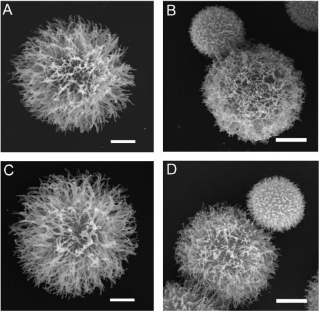
FIG. 3.
Scanning electron microscopy of C. neoformans yeast cells. Cryptococcal cells were grown in SAB medium (ATCC 24067 [A] and H99 [C]) or SAB medium with 0.5 times the MIC of voriconazole (ATCC 24067 [B] and H99 [D]). The yeast cells shown are representative of those seen for each condition. The experiment was performed twice with similar results. Scale bars represent 2 μm.

