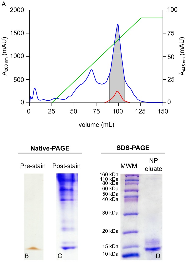
Figure 1. Isolation of a putative CBP by 1D native-PAGE.
E. chloroticus gonad-soluble protein extract was fractionated by anion exchange chromatography, A. Gonad protein extract corresponding to 1 g wet weight gonad was loaded onto a 5 mL HiTrap Q-Sepharose column and bound protein was eluted by a 0–100% gradient of 1M NaCl (green line). The absorbance of the column effluent was monitored at 280 nm (blue) and 445 (red). Fractions absorbing at both 280 nm and 445 nm (grey zone) were pooled concentrated. B. A 20 µL aliquot of the concentrate was analyzed on 1D native-PAGE, shown prior to staining. C. A duplicate loading on 1D native-PAGE was stained with Coomassie blue R-250. D. The yellow/orange band visible on the pre-stained gel in B. was excised and the protein was eluted and then analyzed by 1D SDS-PAGE and stained with Coomassie blue R-250.

