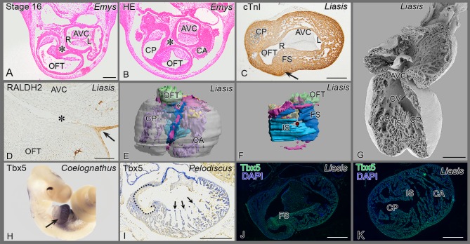
Figure 2. Reptile cardiac development.
(A, B) The epicardial cushion (*) is located between OFT and AV cushions. (C, D) cardiac troponin I (cardiac muscle) and RALDH2 (epicardial cells) stainings show folding septum (arrow, asterisk). (E) 3D reconstruction in an anterior view, the epicardial patches are depicted in pink. See also Figure S1 1 for full animation. (F) right sided view of the septum, folding (FS) and inlet (IS) septum are depicted in shades of blue. For further colors see legend to Fig. 5E. (G) Scanning electron microscopy of anterior inner face, note communication between the three cava. The folding (syn. horizontal) septum is out of view. (H) A sharp decline of Tbx5 mRNA expression (arrow) between cavum dorsale and OFT. (I) Sharp boundary at muscular OFT (inside of dotted line) and wall of cavum pulmonale (outside dotted line), but the tip of trabeculations in the cavum dorsale stain strongly (arrows). (J) Section downstream of Fig C, showing sharp decline of Tbx5 protein expression at folding septum. (K) Section more to the apex of J, showing uniform immunostaining for Tbx5. Abbreviations as in Fig 1, others: AVC: AV cushions; ca, cp, cv: cavum arteriosum, pulmonale and venosum; L left AV orifice; OFT outflow tract cushions; R right AV orifice; →: infolding; * epicardium and EPDCs; • position of cavum venosum in 3D reconstruction of Fig F.

