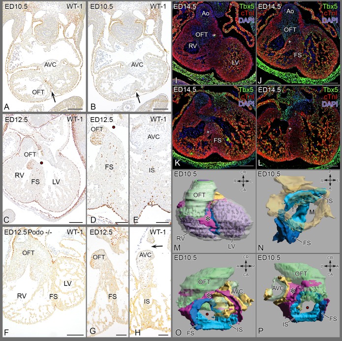
Figure 4. Septum formation in the mouse.
(A, B) Almost transverse sections of the same embryo showing WT1+ epicardial cells in the folding septum (FS,→) at ED 10.5, Fig. B is more apically located. (C, D) epicardial cells in FS of wildtype mouse at ED 12.5 and (E) present in the inlet septum underneath the posterior AV cushion. Note: WT1 staining of mesenchyme in septal OFT cushion is unrelated to epicardial cells. (F-H) Podoplanin mutant with diminutive PEO, presents with sparse epicardium lining the pericardial cavity (F, G) and with an underdeveloped septum lacking EPDCs in both FS (G) and inlet septum (IS) (H). (I-L) Immunostained for Tbx5 in a wild type mouse ED 14.5, four levels from anterior-posterior. (I) Tbx5 in LV trabeculations but not in the RV close to the outflow tract; core of septum is negative. (J-L) More posteriorly located sections, trabeculations in RV belonging to the inlet part become positive for Tbx5. (M-P) Four positions of a 3D Amira reconstruction of ED 10.5. The epicardial cushion in pink (*), the folding septum in dark blue and the inlet septum in light blue. Endocardial cushions in green and the AVC myocardium in yellow. See also Fig. S3 for animated 3D. Abbrev. AVC atrioventricular cushions; FS folding septum; IS inlet septum; LV left ventricle; M mitral orifice; RV right ventricle; OFT outflow tract cushion; T tricuspid orifice, • interventricular communication, + septal band.

