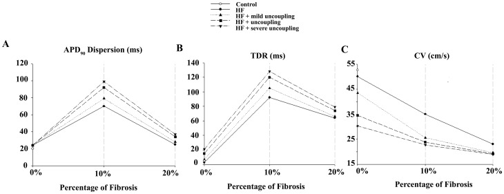
Figure 7. Biomarkers in normal and failing conditions adding structural remodeling with GPB.
Electrophysiological properties measured in a one dimensional transmural ventricular strand under different pathological conditions. The ventricular strands were composed by 82 endocardial and 83 epicardial cells. All simulations were conducted using Grandi et al. (GPB) action potential model [34]. The cases considered were: normal conditions (NC) (white circle), electrical heart failure remodeling (HF) (solid line), electrical heart failure remodeling and mild uncoupling (dotted line), normal uncoupling (dashed line) and severe uncoupling (dotted-dashed line). Different degrees of fibrosis were considered: 10% and 20%. Action potential duration (APD) dispersion (panel A), transmural dispersion of repolarization (TDR) (panel B), and conduction velocity (CV) (panel C) along the whole strand were measured (see Methods section for details).

