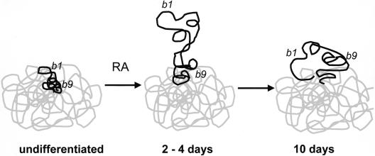
Figure 6.
Changes in chromatin structure at HoxB. In undifferentiated ES cells, the entire Hoxb locus (black) is condensed with the MMU11 CT (gray). However, the 3′ Hoxb1 gene is at the territory surface, poised to respond to retinoic acid (RA). The more 5′ Hoxb9 is further inside the territory. After 2 d of induction with RA, the Hoxb chromatin fiber decondenses, and the 3′ end of the locus, including Hoxb1, is extruded from the CT. By 10 d of differentiation, Hoxb1 is reeled in toward the CT, and Hoxb9 has now left the CT.

