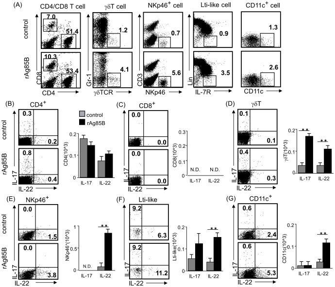
Figure 6. Innate immune cells that secrete Th17-related cytokines are induced by rAg85B administration in BAL fluid.
OVA-immunized (i.p., day0 and 14) and sensitized (5% aerosolized-OVA, day21 to 25) BALB/c mice were challenged with PBS or rAg85B protein (i.p. (100 µg; days 0 and 14) and i.n. (20 µg; days 21, 23, and 25)). At 24 h after the last OVA sensitization, BAL fluid from naïve or OVA sensitized BALB/c mice treated with PBS or rAg85B, were harvested. BAL cells were stimulated with ionomycin and PMA for 5 h, and with brefeldin A added in the last 3 h. Flow cytometry of BAL cells from PBS-treated (upper) and rAg85B protein-treated (lower) OVA-sensitized mice stained with anti-CD3, anti-CD4, anti-CD8, anti-Gr-1, anti-γδ TCR, anti-NKp46, anti-CD11c, anti-CD127 (IL-7R) and Lineage specific marker (CD3, CD19, Gr-1, CD11b, CD11c). Numbers in quadrants indicate percent of cells in each (A). Intracellular IL-17 and IL-22 staining in indicated cells by flow cytometry (dot plots) and absolute numbers of those cell populations (side graphs) in the BAL fluid (B, C, D, E, F, G). Data are representative of at least two independent experiments (**P<0.01 compared with OVA control. error bars, s.d.; n = 6 mice).

