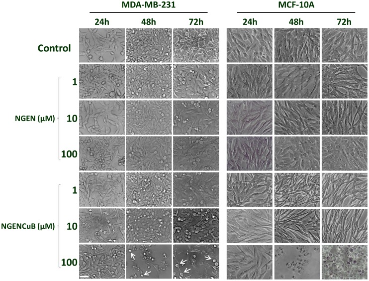
Figure 2. Cellular Morphology of MDA-MB-231 (right panels) and MCF-10A (left panels) control cells, naringenin (NGEN)-treated or naringenin complexed with copper (II) and 2,2′-bipyridine (NGENCuB)-treated cells.
Cells were allowed to grow in a humidified incubator at 37°C in 5% CO2 overnight and then treated with 1, 10 and 100 µM of NGEN or NGENCuB for 24, 48 and 72 hours. Cell morphology was examined under an inverted microscope with amplification of 100×. White arrows indicate detaching round cells. Scale bar = 100 µm.

