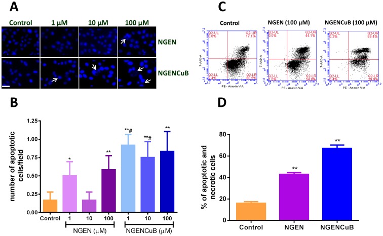
Figure 8. Effects of naringenin (NGEN) or naringenin complexed with copper (II) and 2,2′-bipyridine (NGENCuB) on apoptosis in MDA-MB-231 breast tumor cells.
A. Nuclear 4', 6-diamidino-2-phenylindole (DAPI) staining. Cells treated or not (control) with different concentrations of NGEN or NGENCuB were observed under a fluorescence microscope. Representative phase-contrast and DAPI staining images were taken 24 h post-treatment. B. The number of cells with apoptotic nuclei was counted and plotted in a graphic. Scale bar = 100 µm. C. Cytometry analysis of MDA-MB-231 cells treated or not (control) with 100 µM of NGEN or NGENCuB for 24 hours. After treatment, cells were harvested by trypsinization, centrifuged, washed twice with cold PBS and incubated with PE Annexin V and 7AAD (upper right panel) for 15 minutes in the dark at room temperature and then analyzed by cytometry. D. The percentage of apoptotic and necrotic cells was plotted in a graph. Data represent mean ± SD of three independent assays in triplicate. The results were compared using ANOVA, followed by a Tukey's post-hoc analysis. Asterisks represent *p≤0.05, **p≤0.01 (compared to control), #p≤0.05 (compared to NGEN).

