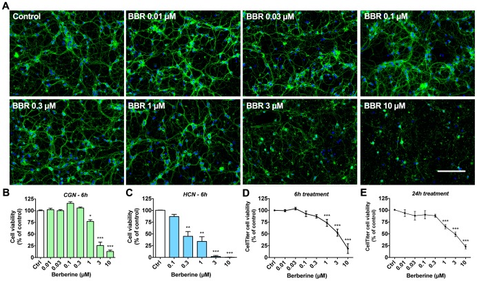
Figure 1. Dose-dependent neurotoxicity of berberine in primary neurons.
Primary neurons (cerebellar granule neurons [CGN; DIV7] and hippocampal neurons [HCN; DIV7]) treated with BBR for 6 hours at concentrations between 0.01 and 10 µM indicate a clear dose-dependent loss of neuronal cell viability with IC50 of roughly 3 µM. (A) CGN were stained for visualization of neurite (β-tubulin III, TUJ1) and nuclear (Hoechst 33342) morphology at 20X magnification. (B) Neuronal cell viability assessed by visual scoring of CGN nuclear morphology after 6-hour treatment with BBR. (C) Neuronal cell viability assessed by visual scoring of HCN nuclei morphology after 6-hour treatment with BBR. (D) Neuronal viability of CGN, as assessed by CellTiter-Glo assay measuring cellular ATP content, for 6-hour treatments with the indicated BBR concentrations. (E) Neuronal viability of CGN, as assessed by CellTiter-Glo assay for 24-hour treatments with the indicated concentrations. For A, green is β-tubulin III, blue is Hoechst; the scale bar represents 100 µm. For panels B, D, E, n = 5. For C, n = 3. For B–E, * = p<0.05, ** = p<0.01, *** = p<0.001.

