Figure 1.
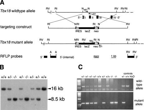
Targeted disruption of the Tbx18 locus. (A) Schematic representation of the targeting strategy. Restriction map of the wild-type locus with boxes representing the first four exons of Tbx18; coding regions are shown in black, noncoding in white. Arrows show location and orientation of PCR primers. Fragments used as RFLP probes are shown. The EcoRV fragment designated as 3′ detects the EcoRI–RFLP shown in B. (A) ApaI; (N) NotI; (RI) EcoRI; (RV) EcoRV; (neo) loxP-flanked neomycin selection cassette; (IRES) internal ribosomal entry site. (B) Southern blot analysis of EcoRI-digested genomic DNA extracted from E18.5 embryos derived from intercrosses of Tbx18/+ mice. Genotypes are indicated above each lane. The 16-kb and 8.5-kb band represent the wild-type and the mutant allele, respectively. (C) PCR genotyping of embryos from heterozygous matings using primers specific for the wild-type (upper panel) and the mutant allele (lower panel), respectively. The genotypes are indicated above each lane.

