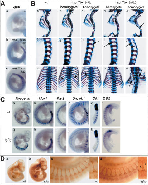
Figure 5.
Ectopic expression of Tbx18 in somites leads to defects in AP-somite polarity and lateral sclerotome development. (A) In situ hybridization analysis of expression of Tbx18-GFP-fusion transcripts in E10.5 transgenic embryos. Embryos hemizygous of msd::Tbx18 line #2 (panel b) and msd::Tbx18 line #20 (panel c) show transgene expression in the PSM, the somitic mesoderm, and the myotome. (B) Skeletal malformations in msd::tbx18 transgenic at E18.5. Genotypes of embryos are indicated above each row. (Panels a–e) Lateral views of whole skeletal preparations of wild-type and transgenic embryos. Arrow in panel e points to reduced pedicles along the vertebral column. (Panels f–j) Higher magnification of the lumbar region of the skeletal preparations shown in panels a–e. Arrows in panels h, i, and j point to reduced pedicles. (Panels k–o) Higher magnification of the rib cage in a dorsal view of the skeletal preparations shown in panels a–e; arrows in l, m, and o point to reduced or missing proximal ribs. (C) In situ hybridization analysis of somite differentiation and polarization in homozygotes of msd::Tbx18 (tg/tg) line #20 at E10.5. Genotypes are indicated in the figure. (Panel g) Myogenin expression is unchanged. (Panel h) Mox1 expression in posterior somite halves is reduced to a thin stripe in the interlimb region of transgenic embryos. (Panel i) The strong posterior Pax9 expression domain is reduced in the interlimb region of msd::Tbx18 transgenic embryos. Anterior is up. (Panel j) Uncx4.1 expression analysis reveals reduction of posterior somite compartments in transgenic embryos. Note that Uncx4.1 expression is normal in the most recently formed five to six somites. (Panel k) Dll1 expression in the PSM and in the most recently formed three somites is unchanged in transgenic embryos, then expression gets progressively weaker. (Panel l) EphrinB2 (E B2) expression is normal in the first four to five somites of transgenic embryos, then it gets weaker. (D) Antineurofilament staining to reveal the organization of spinal nerve projections in homozygotes of msd::Tbx18 line #20 at E10.5. The metameric organization of spinal nerve projections is maintained in transgenic embryos (panel b); however, they appear less bundled (panel d). Higher magnification of the interlimb region of the embryos shown in panels a and b.Anterioristothe left. (Panel d) Arrows in the small inset figure point to colaterals leaving the nerve.

