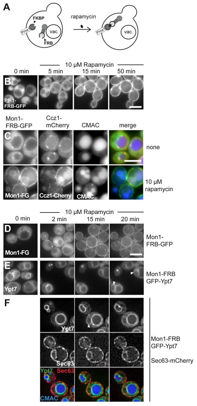
Figure 1. Monitoring of dynamic relocalization of the endosomal GEF complex. (A) Model of rapamycin induced relocalization of tagged proteins to the plasma membrane. (B) Establishment of the rapamycin-induced relocalization. Pfk1 was C-terminally tagged with FRB-GFP in a strain expressing Pma1-FKBP. Cells were monitored without rapamycin (0 min). Then 10 µM rapamycin was added, and cells embedded in agar were monitored for 50 min at 26 °C by fluorescence microscopy. Three time-points are shown. (C) The endosomal GEF complex relocalizes together to the plasma membrane. Mon1-FRB-GFP was expressed from the TEF1 promoter in cells expressing Ccz1-mCherry. Cells were stained with CMAC to label the vacuole and analyzed in the absence and presence of 10 µM rapamycin. Top, cells without rapamycin, bottom, cells after overnight incubation with rapamycin. (D) Time-course of Mon1-relocalization. Mon1-FRB-GFP under the control of the TEF1 promoter was observed in the absence and presence of 10 µM rapamycin at the indicated time points. (E-F) Localization of Ypt7 upon relocalization of Mon1 to the plasma membrane. Mon1-FRB was co-expressed with GFP-tagged Ypt7, and Ypt7 was analyzed by fluorescence microscopy. Analysis was as in (D). White arrows indicate nuclear ER localization of Ypt7. In (F), the same strain also contained Sec63-mCherry to monitor the ER. Cells were stained with CMAC to observe the vacuole, and analyzed in the presence of 10 µM rapamycin.

An official website of the United States government
Here's how you know
Official websites use .gov
A
.gov website belongs to an official
government organization in the United States.
Secure .gov websites use HTTPS
A lock (
) or https:// means you've safely
connected to the .gov website. Share sensitive
information only on official, secure websites.
