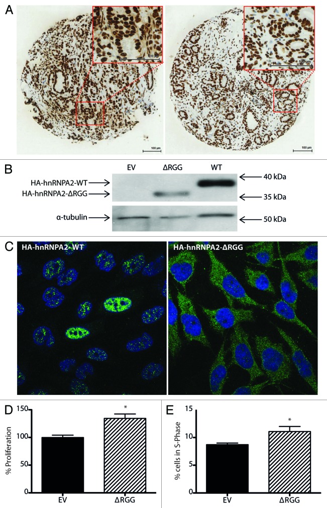
Figure 3. Cytoplasmic hnRNPA2 protein is expressed in clinical PCa and mediates proliferation of PCa cells. (A) Representative images from hnRNPA2-immunostained PCa sections demonstrating cytoplasmic and nuclear protein expression (left panel) and nuclear protein expression alone (right panel). (Bar = 100 µm). (B) Representative western analysis images of PC3 cells transfected with 2 µg of plasmid DNA vectors encoding HA-tagged wild-type (WT) hnRNPA2 or hnRNPA2-ΔRGG or empty vector (EV) control using antibodies to HA tag and α-tubulin. (C) Representative indirect immunofluorescence images of PC3 cells transfected with 2 µg of plasmid DNA vectors encoding HA-tagged WT hnRNPA2 (left) or hnRNPA2-ΔRGG (right) and captured by confocal laser scanning microscopy using indirect immunofluorescence and antibody to HA tag (green) and a DAPI nuclear counterstain (blue). (D) Proliferation of PC3 cells transfected with 0.2 µg of plasmid DNA vector encoding HA-tagged hnRNPA2-ΔRGG was measured using WST-1 proliferation reagent, and normalized to empty vector control. Data from at least three independent experiments with at least five technical replicates were used to calculate the means ± SE (*P = 0.02). (E) Cell cycle distributions of PC3 cells transfected with 2 µg of plasmid DNA vector encoding HA-tagged hnRNPA2-ΔRGG were assessed using flow cytometry. Percentages of cells in the S-phase of cell cycle were estimated from their DNA content as read by propidium iodine. Data from at least three independent experiments were used to calculate the means ± SD (*P = 0.04). (All p-values shown are for comparisons with control conditions).

An official website of the United States government
Here's how you know
Official websites use .gov
A
.gov website belongs to an official
government organization in the United States.
Secure .gov websites use HTTPS
A lock (
) or https:// means you've safely
connected to the .gov website. Share sensitive
information only on official, secure websites.
