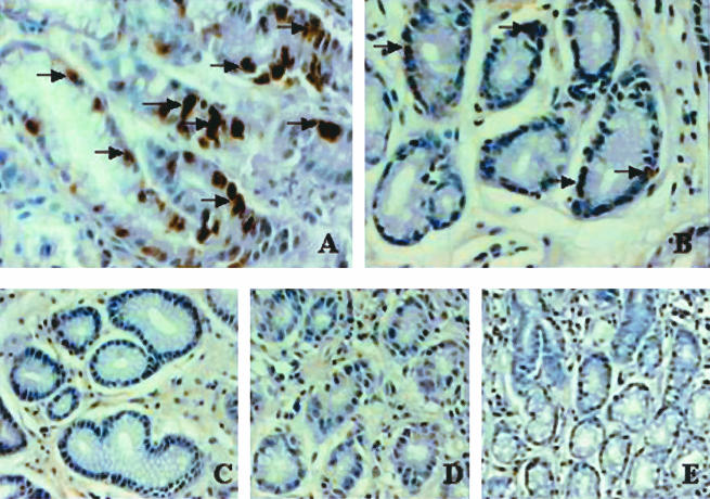
FIG. 5.
Immunohistochemical staining of Egr-1 expression in antral gastric biopsies from H. pylori-infected patients. Immunohistochemical staining was performed on normal biopsies, H. pylori-negative biopsies from patients with mild gastritis, and H. pylori-positive biopsies by the use of an Egr-1 polyclonal antibody (1:50). Patient groups were classified as follows: normal antral-type mucosa, H. pylori negative with mild gastritis (noninfected patients), H. pylori positive with moderate chronic inflammation and focal acute inflammation (H. pylori-infected patients). (A) H. pylori-positive antral gastric biopsy showing increased Egr-1 expression, as indicated by arrows (brown staining). (B) Antral gastric samples from noninfected patients with chronic gastritis showing weak immunostaining (arrows) for Egr-1 expression. (C) Normal antral gastric biopsy showing little or no immunostaining for Egr-1 expression. No primary antibody was used for panel D and an appropriate rabbit polyclonal Ig control was used for panel E. Original magnifications, ×400 (A), ×300 (B and C), and ×200 (D and E).

