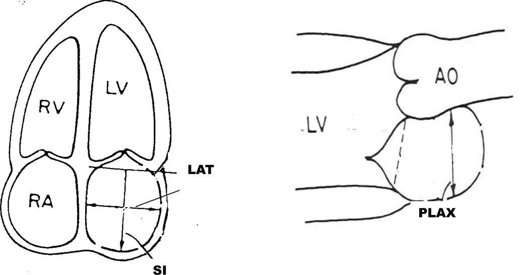
Figure 1.
Schematic representation of the measurement of left atrial volume. The PLAX was taken in the parasternal long axis view. The LAT and SI dimensions were both taken from the apical four chamber view using inner edge to inner edge measurement. LA volume was calculated utilizing the length diameter ellipsoid method, applying the following equation: V = 4π/3 × (PLAX/2) × (LAT/2) × (SI/2).

