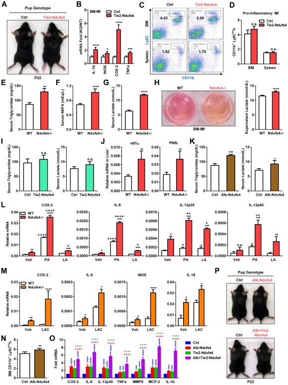
Figure 3. Ndufs4 deletion shifts metabolism to potentiate macrophage activation.
(A) Representative image showing that Tie2-Ndufs4 KO mice had no alopecia.
(B) Expression of inflammatory genes in Tie2-Ndufs4 KO BM-Mfs was increased on day 6 (n=3). (C-D) Percentage of CD11b+Ly6Chi pro-inflammatory macrophages in the bone marrow (BM) and spleen was normal in Tie2-Ndufs4 KO pups on P22. (C) FACS 2D dot plots. (D) Quantification (n=5).
(E-G) Serum triglycerides (E), non-esterified fatty acid (NEFA) (F) and lactate (G) were increased in Ndufs4−/− pups (n=8).
(H) Ndufs4−/− macrophages secrets more lactic acid. Left, images showing the medium of Ndufs4−/− BM-Mf cultures was more acidic (yellow). Right, quantification of lactate in culture medium (n=6).
(I) Serum triglycerides (left) and lactate (right) (n=8).
(J) Expression of Hif1α and Pfkfb was higher in the liver of Ndufs4−/− mice (n=5).
(K) Serum triglycerides (left) and lactate (right) in Alb-Ndufs4 KO mice and controls (n=8).
(L-M) Fatty acids (L) and lactate (M) potentiate the activation of inflammatory genes in Ndufs4−/− macrophages (n=5). BM-Mfs were treated with palmitic acid (PA) or linoleic acid (LA) (400 μM) or lactate (15 mM) for 15 hr.
(N) Percentage of CD11b+Ly6Chi pro-inflammatory macrophages in the bone marrow (BM) was increased in Alb-Ndufs4 KO pups on P22 (n=5).
(O) Expression of inflammatory genes in the spleen was increased in Tie2-Ndufs4 KO and Alb-Ndufs4 KO, and further potentiated in Alb+Tie2-Ndufs4 DKO mice on P22 (n=5).
(P) Alb-Ndufs4 and Alb+Tie2-Ndufs4 DKO mice had no alopecia. Error bars, SD.

