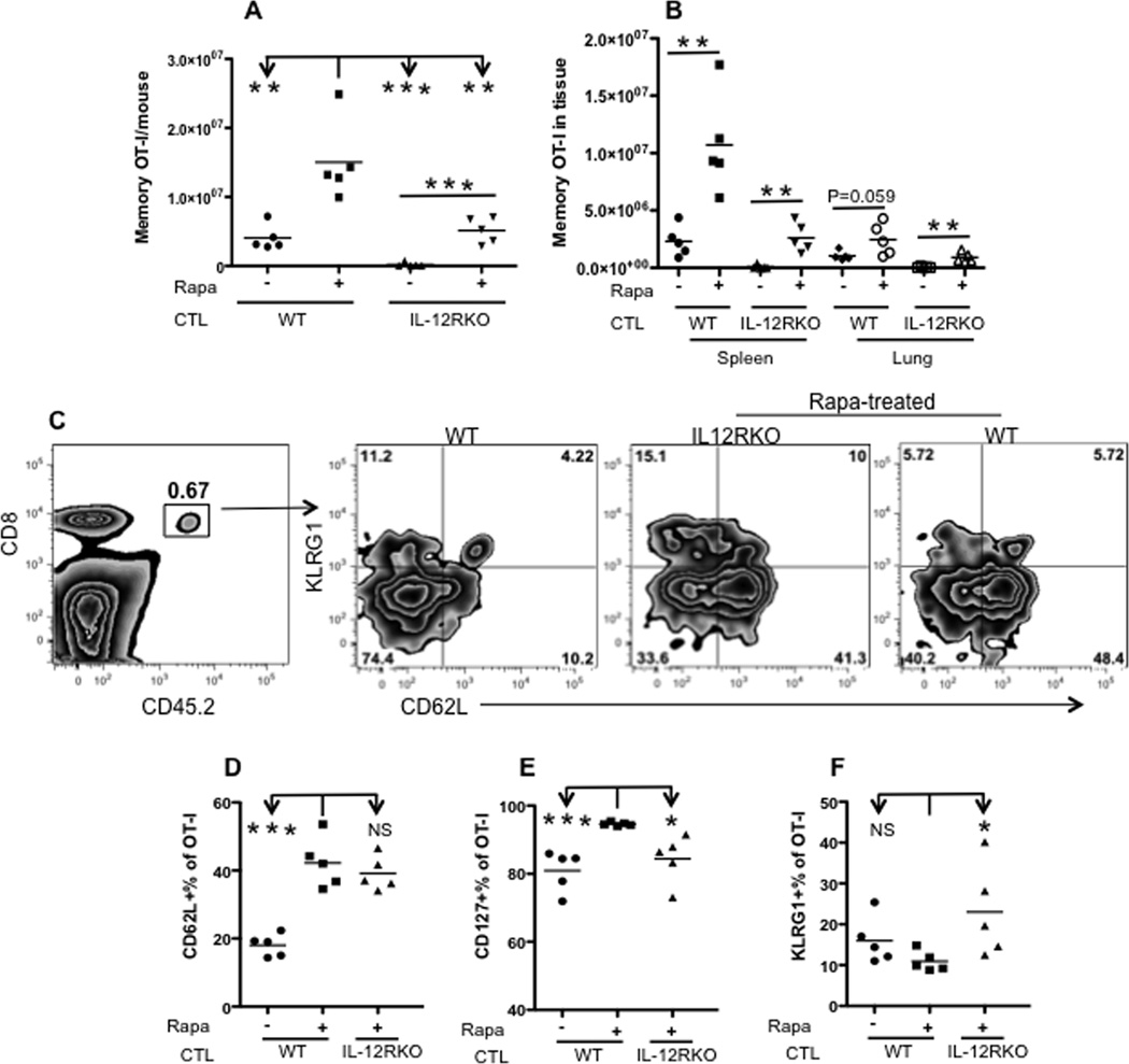
Figure 3. Rapamycin enhances memory CTLs in tissues.
Memory OT-I cells were analyzed in memory mice (similar to those in Fig. 2A) 40 days after VV-OVA infection. A. Comparison of total memory OT-I cells from peripheral lymph nodes, spleen, lung and two sets of femur from each mouse. B. Tissue distribution of memory OT-I cells in spleen and lung. Data were calculated by dividing the number of memory OT-I in one tissue by the number in all examined tissues. C. Representative expression of CD62L/CD127/KLRG1 and corresponding statistics (Student’s t test) (D–F) of memory OT-I cells in spleens from (A). The experiment was repeated three times and similar results were obtained.

