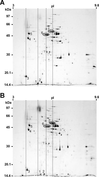
FIG. 2.
Two-dimensional gel electrophoresis of surface protein extract prepared from P. gingivalis W50 (A) and W50CPG (B). Protein extracts (300 μg) were subjected to 2D PAGE, and the gels were stained with colloidal Coomassie blue. The numbered spots were excised, digested, and identified by peptide mass fingerprinting. Spot 1, Kgp14; spot 2, RgpA17; spot 3, RgpA15/Kgp15; spot 4, Kgp39; spot 5, RgpA44; spot 6, RgpA45; spot 7, Kgp48. The dashed lines demonstrate that spots 1, 2, 3, 6 and 7 have moved to a higher pI in the gel from the mutant strain (B). Molecular mass markers are given on the left.

