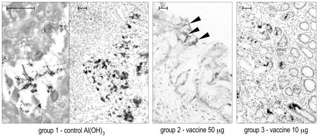
FIG. 5.
Study 1 (weekly immunization): representative IHC results obtained with anti-VacA antibody on 4-μm-thick sections of formalin-fixed, paraffin-embedded gastric antral biopsies at the last sampling (29th week postvaccination). The higher magnification at the left side of the left panel (group 1) shows positively stained H. pylori within a glandular lumen. The solid arrowheads in the central panel indicate some weak, superficial positivity in a dog of group 2. Bar, 50 μm.

