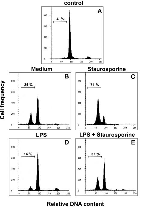
FIG. 1.
Influence of LPS on B-lymphocyte apoptosis. The histograms depict the relative DNA contents of freshly prepared B cells at the onset (control) (A) or of cells recovered after an overnight culture in medium alone (B) or with 10 nM STR (C), 5 μg of S. enterica LPS/ml (D), or LPS plus STR (E). Apoptosis was determined by flow cytometry of 10,000 cells after staining with the DNA probe DAPI. The percentages of apoptotic cells, as determined by the sub-G1 DNA content, are given at the left of each histogram. These data are representative of at least three individual experiments.

