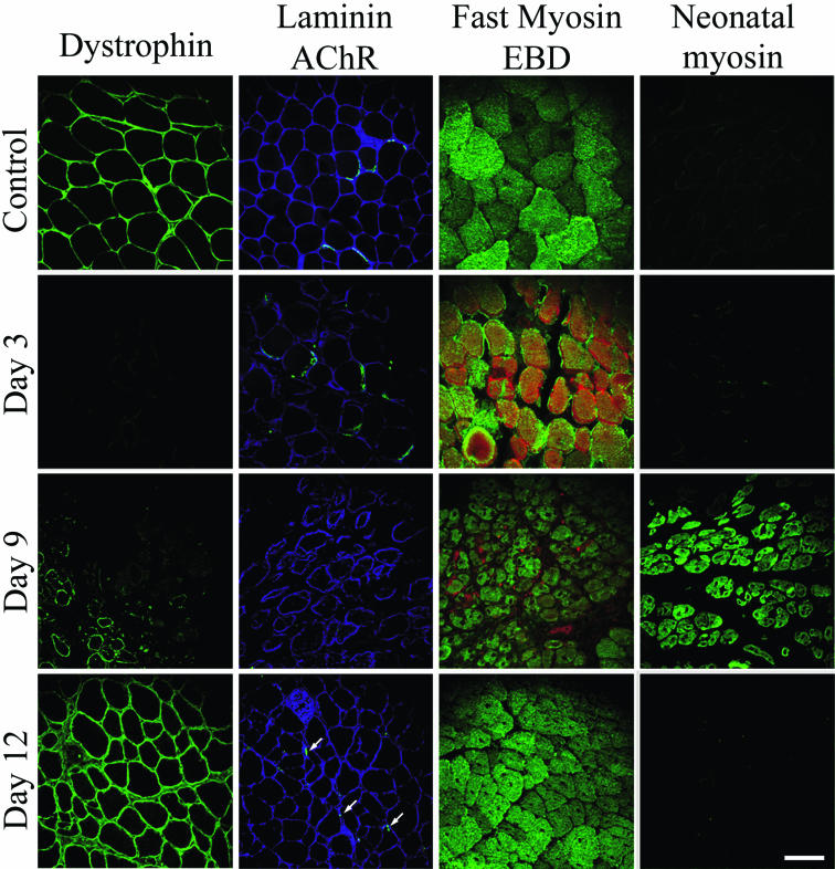
FIG. 4.
Changes in protein expression patterns (revealed by immunostaining of cryosections) during degeneration and regeneration after mice received a sublethal injection of C. sordellii LT into their TA muscles. The immunolabeling of dystrophin, laminin, fast myosin, and neonatal myosin was followed at the times indicated post-LT injection. Staining of acetylcholine receptors (AChRs) was performed with FITC-conjugated α-BTX, and staining with Evans blue dye (EBD) was used to assess muscle membrane injury (red). For details, see Materials and Methods. Note the transient expression of neonatal myosin at day 9 postinjection. Bar = 50 μm.

