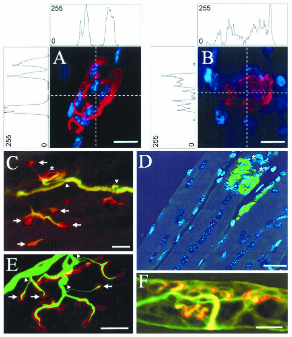
FIG. 6.
Remodeling of the neuromuscular junction in whole-mount LAL muscles obtained from mice at various times after they received a sublethal injection of C. sordellii LT. (A) Control neuromuscular junction in which AChRs are stained with TRITC-conjugated α-BTX (red) and subsynaptic nuclei are stained with DAPI (blue). (B) Staining of AChRs and subsynaptic nuclei (as in panel A) in a junction from a muscle 1 day after LT injection. Note the different intensity profiles for the AChR staining shown in panels A and B, with fluorescence intensity presented as an arbitrary unit of values between 0 and 255 color levels. (C and E) Nodal nerve sprouting (arrowheads) revealed by immunostaining with an antineurofilament antibody (green), and AChRs labeled with TRITC-conjugated α-BTX (red) 30 days post-LT injection. Note the presence of neoformed (arrows) and mature-like AChR clusters (asterisk). (D) Staining of AChRs with FITC-conjugated α-BTX (green), and TOTO-3 dye (blue) staining of nuclei in a regenerated muscle 30 days post-LT injection. Note the presence of large, aligned central nuclei in various fibers. (F) Staining of acetylcholinesterase with TRITC-conjugated fasciculin-2 and axons labeled with an antineurofilament antibody 30 days post-LT injection. Note the similarity between the staining of AChRs in panel C and of acetylcholinesterase in panel F. Bars = 4 μm (A and B), 20 μm (C and D), and 15 μm (E and F).

