Fig. 2.
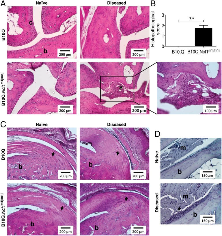
Histology of mannan-induced articular phenotypes. (A) Representative H&E staining of ankle joints of naive/arthritic B10Q (n = 3–5) and B10Q.Ncf1m1j/m1j (n = 3–5) mice after disease initiation (day 4). Microscopic arthritis was absent in diseased B10Q mice, but B10Q.Ncf1m1j/m1j mice had synovial infiltrates (shown as “s”) with mild cartilage damage. (Scale bars: 200 μm. Magnification: 100 μm.) (B) Mean histopathological scores of disease-affected B10Q (n = 5) and B10Q.Ncf1m1j/m1j (n = 7) mouse joints. Data is presented as mean ± SEM. **P < 0.01. (C) Visualization of entheseal inflammation in the Achilles tendon in disease-affected B10Q (n = 5) and B10Q.Ncf1m1j/m1j (n = 7) mice compared with naive mice (arrows). (Scale bars: 200 μm.) (D) Additionally, periostitis was observed in B10Q.Ncf1m1j/m1j mice (n = 7, arrow). (Scale bars: 150 μm.) b, bone; m, muscle; s, synovial membrane.

