Fig. 3.
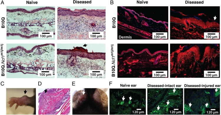
Mannan-induced cutaneous phenotypes in mice. (A) H&E staining of ear tissues from naive and diseased B10Q and B10Q.Ncf1m1j/m1j mice after mannan injection (day 4). (Scale bars: 100 μm.) Dermatitis with intraepidermal accumulation of neutrophilic granulocytes and intracorneal pustules was present in the diseased ears of B10Q mice, whereas mild hyperkeratosis and acanthosis, together with focal parakeratosis and intracorneal pustules (arrow), were observed in the inflamed ears of B10Q.Ncf1m1j/m1j mice. Ears from naive mice did not show any signs of inflammation. (B) Involucrin staining (red) showing abnormal keratinocyte differentiation in the hind paw skin sections of mice after disease induction on day 4 (n = 3 mice per group). Naive mouse skin contained a normal distribution of epidermal keratinocytes. DAPI was used for staining nuclei. (Scale bars: 100 μm.) The white dashed line indicates the border between the epidermal and dermal layers of the skin (white arrow points toward epidermis). (C) Representative image of an “osteogenic nodule” formation (arrow) in a female B10Q.Ncf1m1j/m1j mouse on day 11 after mannan injection. (D) H&E staining of an osteogenic nodule illustrates new bone formation (arrow). (Magnification: 10×). (E) Representative image of ear punching (white arrow). (F) Naive, diseased intact, and diseased-injured ear tissues were stained with MECA-79, depicting dilated blood vessels (arrows) on day 5 after mannan injection (green) (n = 3 mice per group). (Scale bars: 120 μm.)

