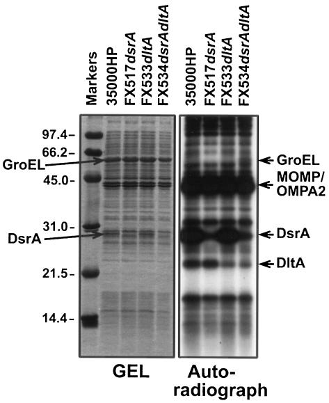
FIG. 8.
Cell surface iodination of whole H. ducreyi cells. Whole cells of H. ducreyi (ca. 5 × 10⁸ CFU) were surface labeled with 125I and subjected to SDS-PAGE in reducing conditions. The gel was stained with Coomassie blue stain, destained, and dried. The left panel of the figure shows the Coomassie blue-stained gel, and the right panel shows the autoradiograph.

