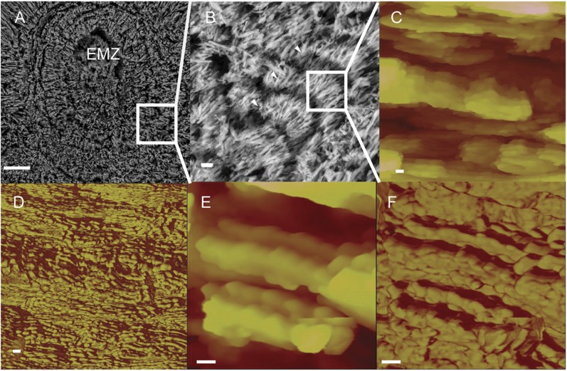
Fig. 2.
Scanning EM images reveal the EMZ and FGSs (white arrowheads) in the S. pistillata skeleton (A and B). AFM of the reticulate structure of the skeleton at increasing magnification reveals the nanoscale building blocks (height images, C and E) and dual composition (phase images, D and F) of skeletal grains. Skeleton growth layers are composed of submicron-sized particles (C and E), which are a composite of an inorganic phase tens of nanometers in diameter (likely aragonite; yellow area in D and F) and an organic matrix phase (red area in D and F). (Scale bars: A, 10 µm; B, 1 μm; C–F, 100 nm.)

