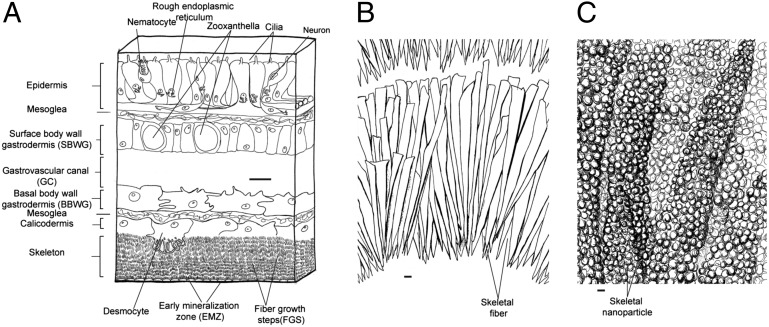
Fig. 4.
Cross-sectional drawing of the histology of coral tissue and skeleton, suggesting a mechanistic understanding of the skeletal growth layers (A). The spatial distribution of SOM proteins are indicated and are suggested to be involved in a diel pattern of skeletal linear extension (night) and skeletal thickening (day) (B). The crystalline units are composed of submicron-sized particles coated with organic matter (C). (Scale bars: A, 10 μm; B, 1 μm; C, 100 nm.)

