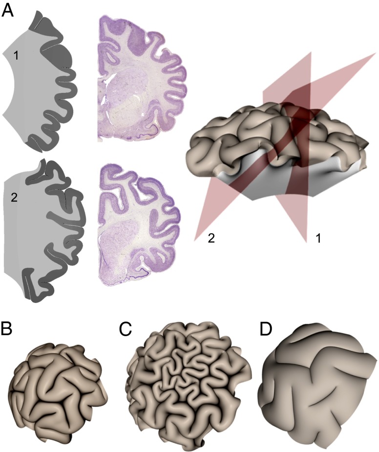
Fig. 4.
(A) Sections of a simulated brain (section planes indicated at right) are compared with coronal sections of a raccoon brain (from www.brainmuseum.org). Cuts through the center of the brain (Upper) and the off the center (Lower) show that we can capture the hierarchical folds but emphasize how misleading sections can be in characterizing the sulcal architecture. (B) Confining our simulations with a uniform pressure of 0.7μ to mimic the meninges and skull leads to a familiar flattened sulcal morphology. (C) Changing the gray matter thickness in a small patch of the growing cortex leads to morphologies similar to polymicrogyria in our simulations. Here g2 = 5 and R/T = 20 except in the densely folded region where R/T = 40. (D) A simulated brain of same physical size as that in C but with a thickened cortex (R/T = 12) and reduced tangential expansion (g2 = 2) displays wide gyri and shallow sulci reminiscent of pachygyric brains.

