Fig. 7.
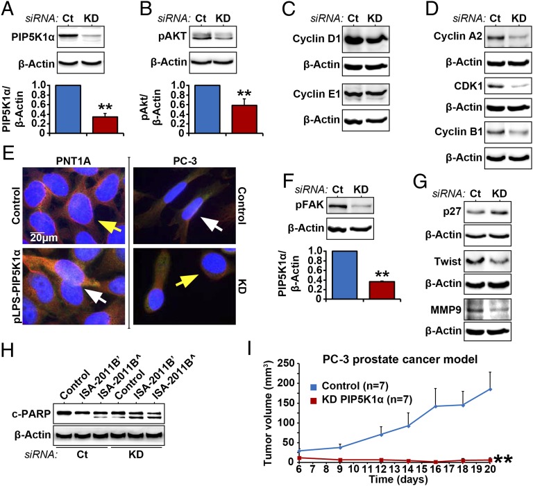
Effect of PIP5K1α inhibition on AKT pathways in PC-3 cells. (A and B) PIP5K1α was depleted by transfecting PC-3 cells with PIP5K1α siRNA (KD) or scramble control (Ct). Immunoblots for PIP5K1α and phosphorylated AKT in PC-3 cells that were transfected with PIP5K1α siRNA (KD) or scramble control (Ct) are shown. Data are presented as average of three independent experiments (±SD). **P < 0.01. (C and D) Immunoblots showing the effect of PIP5K1α knockdown on cyclin D1, cyclin A2, CDK1, and cyclin B1 in PC-3 cells. (E) Morphology of PC-3 cells expressing siRNA to PIP5K1α and PNT1A cells overexpressing PIP5K1α. Antibody to α-tubulin was used for staining. The scale bar is indicated. (F) Immunoblots of phosphorylated FAK in PC-3 cells transfected with PIP5K1α siRNA or scramble control. Data are presented as average of three independent experiments (±SD). Mean expression of phosphorylated FAK in control was 15.30, and in PIP5K1α knockdown was 5.66 (difference = 9.58; 95% CI 4.89–6.42; P < 0.001). (G) Immunoblots of P27, Twist, and MMP9 in PC-3 cells transfected with PIP5K1α siRNA or scramble control. (H) Immunoblots of c-PARP in PC-3 cells treated with vehicle control (DMSO) or ISA-2011B (’20 μM and ^50 μM), with or without PIP5K1α or scramble siRNA cotransfection. (I) s.c. xenografts were established by implanting PC-3 cells expressing control scramble siRNA or PIP5K1α-siRNA into nude mice. The tumors were measured on day 6 after implantation and were followed for a total of 20 d (n = 7 mice per group). Two groups of mice bearing PC3 cells that expressed control siRNA (Control) or PIP5K1α-siRNA (KD-PIP5K1α) are indicated. Difference in tumor growth between the two groups is calculated. **P = 0.008.

