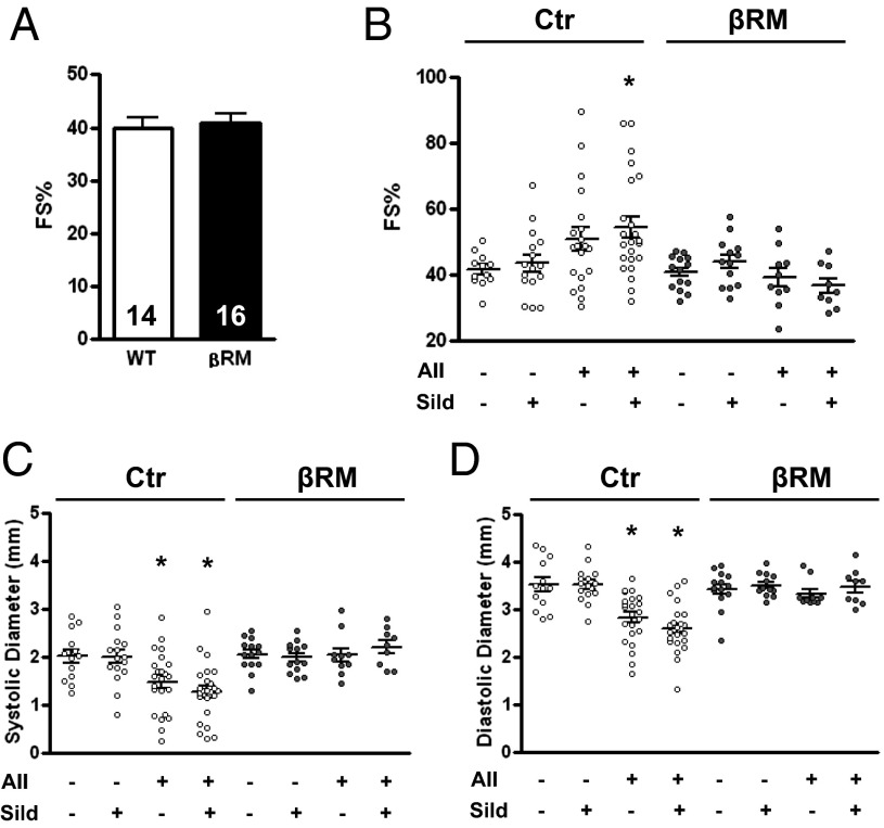
Fig. 2.
Physiological parameters of the animals before and after treatment. Cardiac contractility as measured by echocardiography and expressed as percentage fractional shortening (FS%) in basal conditions (A) and in the experimental groups (B). Left ventricle chamber diameter during systole (C) and diastole (D) (*P < 0.05 vs. corresponding Ctr group, one-way ANOVA with Tukey’s multiple-comparison test).

