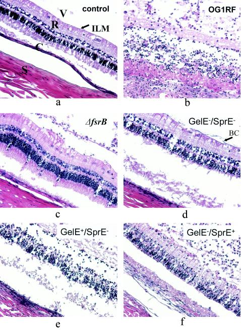
FIG. 3.
Thin-section histopathology (representative slides, hematoxylin and eosin stain). (a) Saline-injected control eye. The vitreous (V) with some extracellular matrix and no inflammatory cells, the internal limiting membrane (arrow labeled ILM), the retina with intact nuclear and plexiform layers (R), the choroid (C), and the sclera (S) can be clearly discerned. (b) Infection with E. faecalis OG1RF after 48 h, showing marked vitreal polymorphonuclear infiltrate, cystoid changes in the ganglionic cell layer, decreased nuclear density of the inner and outer nuclear layers, mild subretinal polymorphonuclear infiltrate, and overall loss of structural integrity. (c) Infection with TX 5266 (OG1RF ΔfsrB) after 48 h, showing mild vitreal polymorphonuclear infiltrate, preserved structure of all retinal layers, and no subretinal inflammatory infiltrate. (d) Infection with TX 5128 (GelE− SprE−) after 48 h. The appearance is similar to that after infection with TX 5266 (OG1RF ΔfsrB). Note the bacterial clusters on the internal limiting membrane (arrow labeled BC). (e) Infection with TX 5243 (GelE+ SprE−) after 48 h. The appearance is similar to that after infection with the wild type, OG1RF. (f) Infection with TX 5264 (GelE− SprE+) after 48 h, with marked vitreal inflammation, mild to moderate retinal and subretinal infitration, and relatively preserved retinal structure.

