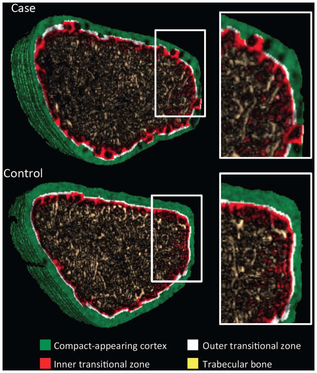
Figure 1.
Representative segmented image obtained at the ultradistal radius using non threshold-based image analysis in a postmenopausal women with (Case) and without (Control) forearm fracture. The full cross section and the magnified image show the presence of porosity within the compact appearing cortex (green) and the outer (white) and inner (red) transitional zones, and loss of trabecular bone (yellow) in the case, and less so in the control.

