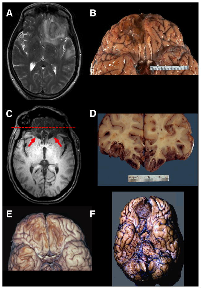
Figure 4. Lesions to Orbitofrontal Cortex.
(A) A T2 axial MRI image of an acute right OFC contusion resulting from trauma (accidental fall down stairs). (B) A ventral view of a postmortem specimen showing chronic residual loss of tissue in right OFC. Note also the damage to the olfactory bulb. (C)A T1 axial MRI image showing extensive bilateral damage to the OFC (red arrows). (D) Acoronal post-mortem slice of a patient with fatal traumatic brain injury due to extensive OFC contusions. If this patient had survived, his/her MRI image would resemble (C). The red dashed line on the MRI in (C) shows the approximate site of this post-mortem coronal slice. (E) A ventral view of a post-mortem specimen showing chronic bilateral loss of tissue in the OFC due to trauma. (F) A post-mortem specimen (ventral view) of an unsuspected right meningioma in a patient with behavioral changes. Neuropathological specimens in (B) and (D) are compliments of Professor Edward C. Klatt, University of Utah (http://library.med.utah.edu/WebPath/CNSHTML/CNSIDX.html). Neuropathological specimen in (E) is compliments of Professor Walter Finkbeiner, UC San Francisco.

