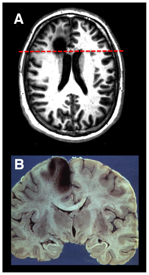
Figure 5. Lesions to Medial Prefrontal Cortex.
(A) An axial T1 MRI image of a patient with a chronic left medial frontal lobe lesion due to the resection of a low-grade glioma. (B)A coronal neuropathological specimen of an infarct in the left medial PFC (note the hemorrhagic conversion in the cortical mantle). The dashed red line on the MRI in (A) shows the approximate site of the post-mortem coronal slice. Image in (A) is compliments of Professor Marianne Løvstad, University of Oslo. Neuropathology specimen in (B) is compliments of Professor Dimitri Agamanolis, Akron Children’s Hospital (http://neuropathology-web.org/).

