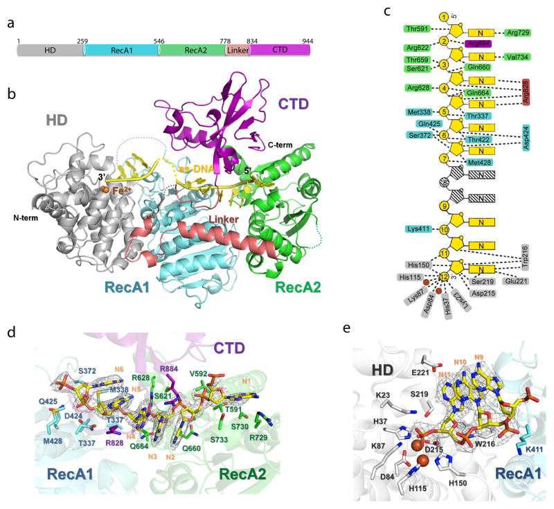
Figure 1. Overall structure of ss-DNA-bound T. fusca Cas3 protein.
(a). Domain organization and (b) overall structure. HD, RecA1, RecA2, linker, and CTD domains and the bound ss-DNA substrate are colored in silver, cyan, green, pink, magenta and yellow, respectively. This coloring scheme is followed throughout figures. (c) DNA recognition diagram. Contacts from Cas3 residues to base, ribose, or phosphate of ss-DNA are illustrated in dashed lines. Two disordered nucleotides are shown in stripes. Views of the Cas3-DNA interaction in the helicase (d) and HD nuclease (e) regions. 2Fo-Fc ss-DNA electron densities (1.5σ) in light grey, protein in cartoon, and DNA-contacting side chains in sticks.

